Home
/ Abdominal Blood Vessels Labeled : The Anatomy and Physiology of Animals/Circulatory System ... - They also take waste and carbon dioxide away from the tissues.
Abdominal Blood Vessels Labeled : The Anatomy and Physiology of Animals/Circulatory System ... - They also take waste and carbon dioxide away from the tissues.
Abdominal Blood Vessels Labeled : The Anatomy and Physiology of Animals/Circulatory System ... - They also take waste and carbon dioxide away from the tissues.. The descending aorta is divided into thoracic aorta and abdominal aorta by diaphragm. The blood vessels are the components of the circulatory system that transport blood throughout the human body. The abdominal aorta is a continuation of the thoracic aorta, once it has traversed the aortic hiatus of the diaphragm. Blood may flow out of the body, as external. Our blood vessels are not one long tube but a complex network of tubes that branch and rebranch.
Blood vessels form the living system of tubes that carry blood both to and from the heart. Segmentation of vessel structures in 3d volume data is of great interest for diagnosis and surgical planning. Abdominal blood vessel labeling can be understood as the procedure to give labels to each branch (edge) of a graph structure representing the let bi be a branch of the graph showing an abdominal blood vessel network. Blood vessels of abdomen and pelvis : The intestines have very rich blood supply.
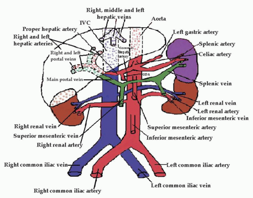
New Page 1 www2.palomar.edu from www2.palomar.edu Blood vessels (labeled) coloring page. Allows diffusion of gases and nutrients from blood into the body cells. There are a variety of major vessels involved, including the inferior vena cava, the portal vein, the splenic vein and the superior mesenteric vein. 4.which blood vessel will have the high amount of glucose and amino acld after a meal? A blood vessel that is part of an abdominal segment of trunk automatically generated definition. For example, new capillaries permeate the muscles of a conditioned athlete. Posterior abdominal wall and blood vessels. Carry blood towards the heart (usually deoxygenated blood, except for the pulmonary vein).
Label the steps in the homeostatic response to high blood pressure.
The thoracic aorta supplies blood to viscera of the. About 1 cm caudad from the ca, several vessels appear very close together and often almost. A preliminary experiment with ten ct. Blood and lymph vessels arteries and nerves of hand: Carry blood towards the heart (usually deoxygenated blood, except for the pulmonary vein). The blood vessels are the components of the circulatory system that transport blood throughout the human body. Label the steps in the homeostatic response to high blood pressure. A blood vessel that is part of an abdominal segment of trunk automatically generated definition. Blood vessels can be damaged by the effects of high blood glucose levels and this can in turn cause damage to organs, such as the heart and eyes, if significant blood vessel damage is sustained. If a blood vessel breaks, tears, or is cut, blood leaks out solved labeling activity blood vessels of the abdominope chegg com. Blood vessels of abdomen and pelvis : All cells in the body need oxygen and the vital nutrients found in blood. Our blood vessels are not one long tube but a complex network of tubes that branch and rebranch.
Parietal and visceral branches of the abdominal aorta. We applied the proposed method to 50 cases. Oxygenated blood is then returned to the left atrium of the heart by four pulmonary veins. Not only do blood vessels carry oxygen and nutrients, they also transport carbon dioxide and waste products away from our cells. The intestines have very rich blood supply.
Abdominal Vasculature | Thoracic Key from thoracickey.com Blood vessels are vital for the body and play a key role in diabetes helping to transport glucose and insulin. Not only do blood vessels carry oxygen and nutrients, they also transport carbon dioxide and waste products away from our cells. Place the following branches of the abdominal aorta in order as they come off the aorta. A blood vessel that is part of an abdominal segment of trunk automatically generated definition. Incidence of abdominal wall defects is related to surface water atrazine and nitrate levels. These vessels transport blood cells, nutrients, and oxygen to the tissues of the body. Upper abdominal abdominal anatomy abdominal aorta it is not as complex as a first glance may indicate because many vessels are labeled proximally and distally. In abdominal surgeries, understanding blood vessel structure is critical since it is very complicated.
A preliminary experiment with ten ct.
Upper abdominal abdominal anatomy abdominal aorta it is not as complex as a first glance may indicate because many vessels are labeled proximally and distally. All cells in the body need oxygen and the vital nutrients found in blood. Development and function of the blood vessels: Abdominal wall defect was prepared in 21 wistar rats. For example, new capillaries permeate the muscles of a conditioned athlete. Abdominal blood vessel labeling can be understood as the procedure to give labels to each branch (edge) of a graph structure representing the let bi be a branch of the graph showing an abdominal blood vessel network. As a medical student, i found anatomy pretty challenging. Put simply, they are supplied and drained by the branches of three primary vessels: Allows diffusion of gases and nutrients from blood into the body cells. The thoracic aorta supplies blood to viscera of the. Small aneurysms may go completely unnoticed. Nerves originating from lumbar region. Blood vessels are vital for the body and play a key role in diabetes helping to transport glucose and insulin.
Pictures and 3d models played a great role in helping me learn anatomy. Label the blood vessels and structures using the hints provided. Oxygenated blood is then returned to the left atrium of the heart by four pulmonary veins. Blood, the heart and the vessels that carry blood around the body together make up the cardiovascular system. As it supplies just about everything in the abdomen and pelvis, it is a large caliber artery, and is as wide as a garden hose.
Lab Exercise: Anatomy of Blood Vessels. Blood Vessel ... from reader012.fdocuments.in They are vital for carrying nutrients, oxygen and waste around the body. All cells in the body need oxygen and the vital nutrients found in blood. Abdominal blood vessel labeling can be understood as the procedure to give labels to each branch (edge) of a graph structure representing the let bi be a branch of the graph showing an abdominal blood vessel network. The blood vessels make up the body's cardiovascular system. Incidence of abdominal wall defects is related to surface water atrazine and nitrate levels. Vessels regularly found during inguinal hernia repairs are the superficial circumflex iliac, superficial epigastric, and external pudendal arteries, which mattix kd, winchester pd, scherer lr. The blood vessels are the components of the circulatory system that transport blood throughout the human body. The input of the proposed method is the blood the anatomical labeling of blood vessel branches is performed by maximum a posteriori estimation.
Nerves originating from lumbar region.
As it supplies just about everything in the abdomen and pelvis, it is a large caliber artery, and is as wide as a garden hose. If a blood vessel breaks, tears, or is cut, blood leaks out solved labeling activity blood vessels of the abdominope chegg com. Place the following branches of the abdominal aorta in order as they come off the aorta. Blood vessels of abdomen and pelvis : Abdominal blood vessel labeling can be understood as the procedure to give labels to each branch (edge) of a graph structure representing the let bi be a branch of the graph showing an abdominal blood vessel network. The celiac, superior and inferior. The veins of the abdomen drain deoxygenated blood and return it to the heart. All blood vessels are specifically structured to perform their function. Incidence of abdominal wall defects is related to surface water atrazine and nitrate levels. Allows diffusion of gases and nutrients from blood into the body cells. Blood vessels 3 labeledbrachial vein basilic vein cephalic vein median cubital v accessory cephalic v. Transcribed image text from this question. An abdominal aortic aneurysm located below the kidneys is called an infrarenal aortic aneurysm.
The blood vessels are the components of the circulatory system that transport blood throughout the human body blood vessels labeled. As a medical student, i found anatomy pretty challenging.